
TL;DR

Turn your wind-down time into listening time
- Thousands of audiobooks, podcasts and originals
- Listen on your phone, tablet or Echo — also offline
- Cancel anytime
In an interview with Being Patient, Columbia radiologist Michael Lipton described research into quantitative MRI methods that measure brain features not readily visible on routine scans. He said a standard MRI reported as normal can still miss microscopic or network-level changes; the interview does not establish that quantitative MRI is a routine diagnostic test or can determine the cause of an individual’s symptoms.
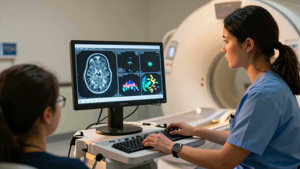
Being Patient has published an interview with Columbia University radiologist Michael Lipton on how quantitative MRI research may detect and measure subtle brain changes that a routine scan can miss. Lipton said a report describing a conventional MRI as normal means that the radiologist did not see an abnormality in the images—not that microscopic brain changes or every possible cause of neurological symptoms have been ruled out.
Lipton, a professor of radiology at Columbia University Irving Medical Center and an attending radiologist at NewYork-Presbyterian, described conventional brain MRI as an examination of images interpreted by a radiologist. Those scans can identify visible problems such as a sizable stroke or brain tumor, he said. But the assessment depends on what can be seen in the images, and some disease-related changes occur at a smaller scale.
The interview discusses quantitative MRI as a research approach for measuring brain features rather than relying only on a visual reading. Lipton said the techniques he studies may reveal subtle changes, including brain volume loss associated with Alzheimer’s disease. He also discussed research on repetitive head impacts in soccer players. The supplied interview material does not give specific study results, participant numbers, or performance measures for these methods.
Lipton said persistent symptoms can occur despite an MRI that appears unremarkable. He distinguished large, visible injuries from problems involving higher functions such as memory, attention, personality, sleep, or balance, which may relate to the way brain cells and networks function. He described microscopic connections between nerve cells as one reason such changes may not appear on a standard clinical scan.
What a Normal Scan Can Miss
The distinction matters to people who receive a normal MRI result but continue to have symptoms. A routine report describes what was visible on that examination; it does not, by itself, explain every symptom or rule out all biological changes. Lipton’s comments also caution against treating a normal scan as a general declaration that the brain is healthy.
Quantitative methods could give researchers ways to measure subtle changes over time or compare brain features across groups. That may support research into conditions involving memory, injury, or neurodegeneration. However, the interview describes research potential, not proof that these tools can diagnose a particular disease in an individual or replace a clinician’s assessment.
brain MRI anatomy and neuroscience textbook
As an affiliate, we earn on qualifying purchases.
As an affiliate, we earn on qualifying purchases.
From Visual Reads to Measurements
Routine MRI produces multiple images of the brain that a radiologist reviews for visible abnormalities. As Lipton explained, that visual interpretation can be useful for detecting structural problems once they are large enough to show on the scan. It is not the same as examining tissue under a microscope, where smaller changes may be apparent.
Lipton’s work includes advanced quantitative imaging and studies of concussion and repeated head impacts in soccer players. The interview links that research to a broader limitation of conventional imaging: brain function depends on cells and connections that may be too small to distinguish in standard pictures. The article presents these points as an explanation of the research, not as a new clinical guideline or a report of a newly approved test.
“That does not necessarily mean that the brain is normal.”
— Michael Lipton, professor of radiology at Columbia University Irving Medical Center, in an interview with Being Patient
Limits of the Evidence Presented
The supplied interview does not specify which quantitative MRI protocols were discussed, how accurately they detect particular changes, or whether the methods are available in routine patient care. It also provides no study sample sizes, numerical findings, or comparisons with other diagnostic tools. Claims about possible detection of subtle volume loss and changes linked to disease should therefore be understood as Lipton’s description of research, not as established diagnostic performance.
The interview also does not determine why a particular person has ongoing symptoms after a normal scan. A routine MRI result alone cannot settle that question, and the article does not offer a diagnostic pathway or patient-specific medical advice.
Research and Clinical Validation
The next steps depend on the underlying studies: researchers would need to report how quantitative measures perform, which conditions and populations they apply to, and whether results can be reproduced. Further evidence would also be needed to establish how such measurements might complement standard imaging and clinical evaluations.
Being Patient’s interview offers an account of Lipton’s research and interpretation, but it does not announce a trial, regulatory decision, or change in clinical practice. Readers with ongoing neurological symptoms should discuss their concerns and test results with a qualified health professional, who can interpret them in the context of their history and examination.
Key Questions
Does a normal brain MRI prove that the brain is healthy?
No. Lipton said a normal report means the radiologist did not see an abnormality in the images reviewed. It does not rule out every microscopic change or explain every possible symptom.
What does quantitative MRI do?
As described in the interview, quantitative MRI research uses measurements of brain features to look for changes that may not be apparent through visual inspection of routine images. The supplied material does not identify a specific protocol or establish its diagnostic accuracy.
Can quantitative MRI diagnose Alzheimer’s disease?
The interview says quantitative techniques may detect subtle brain volume changes associated with Alzheimer’s disease. It does not establish that the methods can diagnose Alzheimer’s in an individual or replace other clinical assessments.
Why can symptoms persist after an unremarkable scan?
Lipton said some symptoms may relate to microscopic structures or communication across brain networks that are not visible on a routine MRI. The interview does not identify the cause of symptoms in any particular person.
Is quantitative MRI a routine clinical test?
The supplied article describes Lipton’s research but does not say that these methods are routinely available or used to guide standard patient care.
Source: rss
Halloween Picks
halloween
As an affiliate, we earn on qualifying purchases.